 anotemy
Sign in
Try it free
Femur — Anatomy Notes, Diagram & Quiz
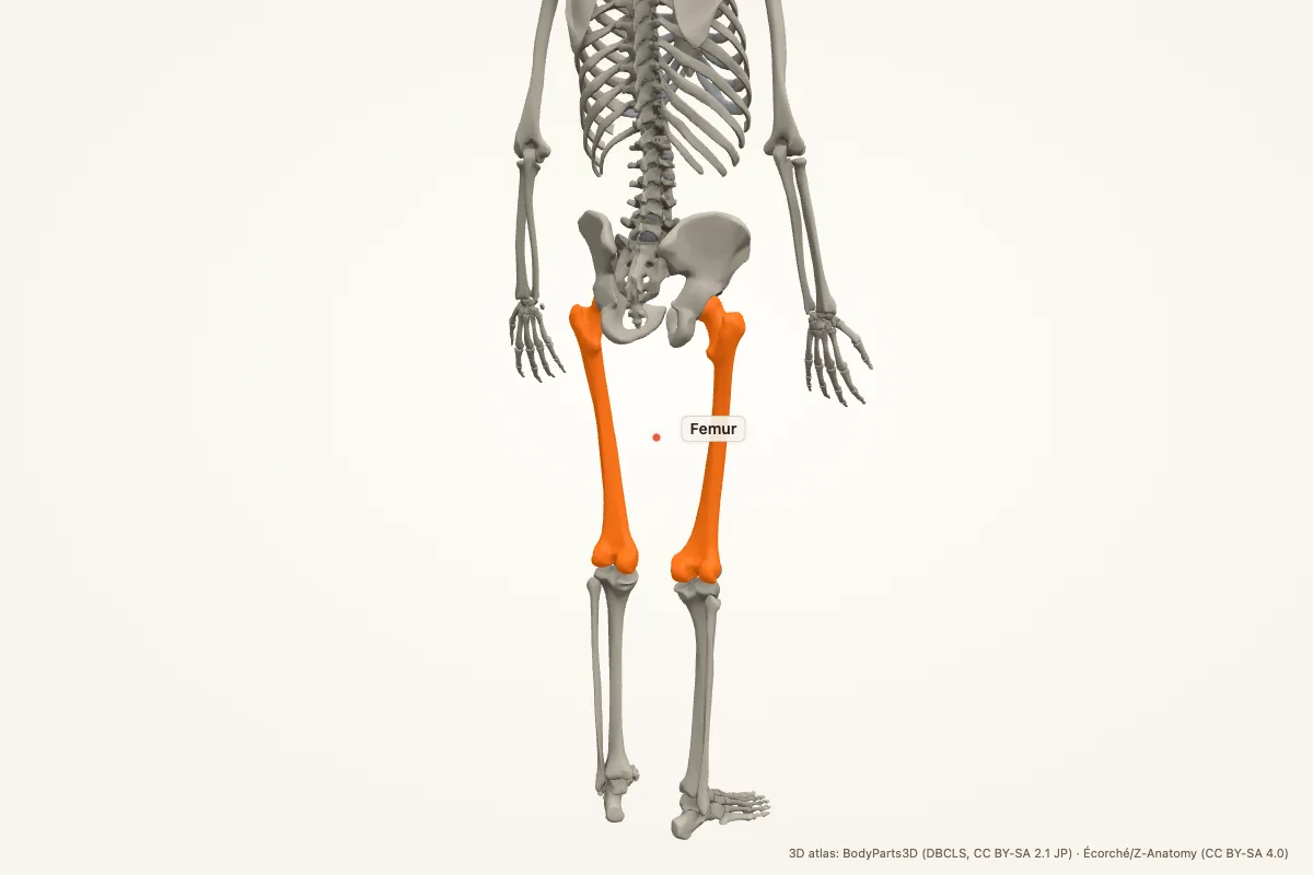
The femur is the thigh bone and the longest, strongest bone in the body. It articulates with the acetabulum at the hip and the tibia and patella at the knee. Its head, neck, trochanters, shaft and condyles are key exam landmarks.
Key facts
| Region | Thigh; hip to knee |
|---|---|
| Proximal landmarks | Head, fovea capitis, neck, greater and lesser trochanters, intertrochanteric line and crest |
| Shaft | Linea aspera, gluteal tuberosity, adductor tubercle |
| Distal landmarks | Medial and lateral condyles, epicondyles, intercondylar fossa, patellar surface |
| Articulations | Hip (acetabulum), knee (tibia and patella) |
| Key blood supply | Medial circumflex femoral artery to the head and neck |
| Main muscles | Gluteals, iliopsoas, quadriceps, hamstrings, adductors |
| Clinical | Femoral neck fracture, intertrochanteric fracture, shaft fracture, avascular necrosis |
Where is the femur located?
The femur is the bone of the thigh. It runs from the hip joint to the knee joint and is the longest and strongest bone in the body. The head points medially into the acetabulum; the shaft angles slightly medially toward the knee, so the knees sit closer to the midline than the hips.
The femur is covered by thick muscles anteriorly, posteriorly and medially. Its neck is a common fracture site, especially in older adults with osteoporosis, because the neck transmits body weight through a narrow bridge of bone.
Proximal femur
The head is spherical and covered by articular cartilage except at the fovea, where the ligament of the head attaches. The neck connects the head to the shaft at an angle. The greater trochanter is lateral and palpable; the lesser trochanter is posteromedial and is the insertion of iliopsoas.
The intertrochanteric line runs anteriorly between the trochanters; the intertrochanteric crest runs posteriorly. The neck is intracapsular, which matters clinically: fractures here can disrupt the medial circumflex femoral artery and cause avascular necrosis of the head.
Femoral shaft
The shaft is roughly cylindrical and has a prominent posterior ridge, the linea aspera. The linea aspera gives attachment to the adductors, vasti and short head of biceps femoris. Proximally, the gluteal tuberosity receives the gluteus maximus; distally, the linea aspera divides into the supracondylar lines.
The shaft has a nutrient foramen and is surrounded by a strong periosteum. Femoral shaft fractures can bleed heavily because the bone is large and the surrounding muscles are well vascularised.
Distal femur
The distal femur expands into medial and lateral condyles, separated posteriorly by the intercondylar fossa. The patellar surface lies anteriorly between the condyles. The medial and lateral epicondyles sit above the condyles and give attachment to collateral ligaments.
The adductor tubercle sits on the medial epicondyle and receives the adductor magnus. The popliteal surface is posterior and forms the floor of the popliteal fossa. The condyles articulate with the tibial plateau and the patella.
Clinical relevance
- Femoral neck fracture: common in osteoporosis; risks avascular necrosis of the femoral head.
- Intertrochanteric fracture: extracapsular, often with more bleeding and a different fixation strategy.
- Femoral shaft fracture: can cause significant blood loss and fat embolism.
- Avascular necrosis: loss of blood supply to the femoral head causes pain and collapse.
- Distal femoral fracture: can involve the knee joint and popliteal vessels.
- Hip fracture: the leg may appear shortened and externally rotated.
Memory aid: neck, trochanters, condyles
Head → neck → greater trochanter (lateral) → lesser trochanter (posteromedial) → shaft → condyles.
The medial circumflex femoral artery supplies most of the femoral head. That is why a displaced neck fracture threatens avascular necrosis.
Femur anatomy quiz
Five questions. Open each answer after you commit to a response.
-
What is the longest bone in the body?
Reveal answer
The femur.
-
Which trochanter is lateral and palpable?
Reveal answer
The greater trochanter.
-
Which muscle inserts on the lesser trochanter?
Reveal answer
Iliopsoas.
-
What is the posterior ridge of the femoral shaft called?
Reveal answer
The linea aspera.
-
Which artery supplies most of the femoral head?
Reveal answer
The medial circumflex femoral artery.
Common questions
Where is the femur located?
The femur is the thigh bone, running from the hip joint to the knee joint. Its head sits in the acetabulum, and its distal condyles articulate with the tibia and patella. It is the longest and strongest bone in the body.
What are the key landmarks of the femur?
Proximally: head, fovea, neck, greater and lesser trochanters, intertrochanteric line and crest. The shaft has the linea aspera. Distally: medial and lateral condyles, epicondyles, intercondylar fossa and patellar surface.
Why is a femoral neck fracture dangerous?
The femoral head receives most of its blood from the medial circumflex femoral artery, which runs along the neck. A displaced neck fracture can tear those vessels and cause avascular necrosis of the head, so fixation or replacement is often needed.
Which muscles attach to the femur?
The gluteals attach to the greater trochanter and gluteal tuberosity; iliopsoas inserts on the lesser trochanter; the quadriceps and hamstrings attach around the linea aspera and condyles; the adductors attach along the shaft and adductor tubercle.
What is the linea aspera?
The linea aspera is the prominent posterior ridge of the femoral shaft. It gives attachment to the adductors, vasti and short head of biceps femoris, and it divides distally into the supracondylar lines.
How does a hip fracture present?
A hip fracture often presents with pain, inability to bear weight, and a shortened, externally rotated leg. The exact deformity depends on whether the fracture is intracapsular or extracapsular. Urgent imaging and orthopaedic assessment are required.
Sources
- Standring, S. (ed.). Gray’s Anatomy, 42nd edition. Elsevier, 2020.
- TeachMeAnatomy — Femur
- Kenhub — Femur
- StatPearls / NCBI Bookshelf — femur anatomy search
3D atlas and image credits
Anatomy models: BodyParts3D, © Database Center for Life Science (DBCLS), CC BY-SA 2.1 Japan. Écorché by Charleeh, derived from BodyParts3D and Z-Anatomy, CC BY-SA 4.0. Rendered in anotemy. Changes: highlighting, framing, labels and background removal. Images: CC BY-SA 4.0.
The 3D models are third-party assets under Creative Commons share-alike licences. They keep those licences; the page text is separate. If you reuse an image, keep the attribution and the share-alike licence.