 anotemy
Sign in
Try it free
Quadriceps — Anatomy Notes, Diagram & Quiz
The quadriceps femoris is a group of four muscles on the front of the thigh: rectus femoris, vastus lateralis, vastus medialis and vastus intermedius. They extend the knee; rectus femoris also flexes the hip. All are innervated by the femoral nerve (L2–L4).
Key facts
| Region | Anterior thigh |
|---|---|
| Muscles | Rectus femoris, vastus lateralis, vastus medialis, vastus intermedius |
| Origin | Rectus femoris: anterior inferior iliac spine; vasti: femoral shaft |
| Insertion | Patella via quadriceps tendon, then tibial tuberosity via patellar ligament |
| Action | Knee extension; rectus femoris also flexes the hip |
| Innervation | Femoral nerve (L2–L4) |
| Blood supply | Femoral artery and descending genicular artery |
| Clinical | Patellar tendon reflex, quadriceps atrophy, patellofemoral pain, tendon rupture |
Where is the quadriceps located?
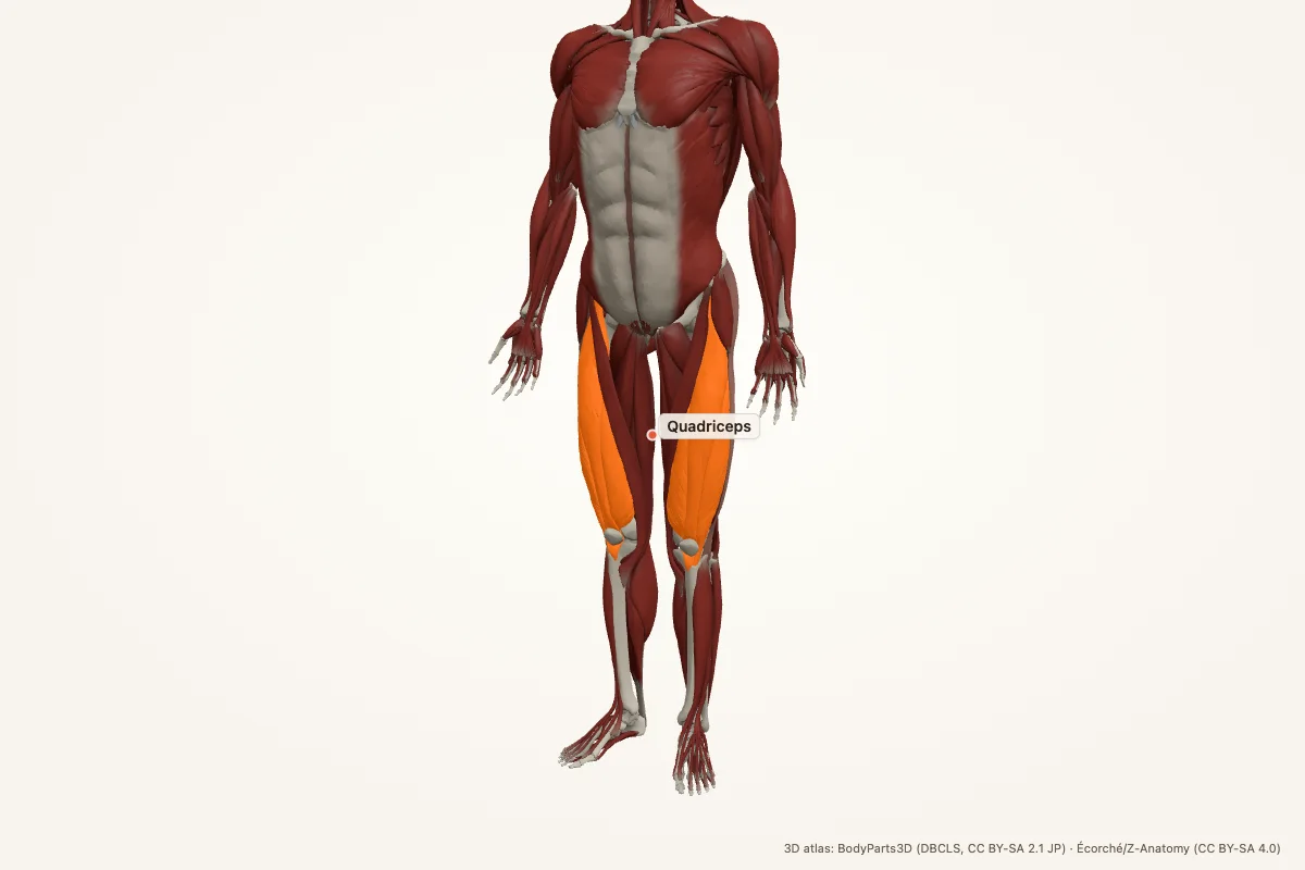
The quadriceps femoris occupies the anterior compartment of the thigh. It forms the bulk of the front of the thigh and converges on the patella. The four muscles are the rectus femoris, vastus lateralis, vastus medialis and vastus intermedius.
The rectus femoris is superficial and central; the vastus lateralis is on the lateral side; the vastus medialis is on the medial side and forms the medial bulge above the knee; the vastus intermedius lies deep to rectus femoris.
Origin and insertion
Rectus femoris arises from the anterior inferior iliac spine and the superior rim of the acetabulum. The vastus lateralis arises from the greater trochanter and linea aspera; vastus medialis from the intertrochanteric line and medial linea aspera; vastus intermedius from the anterior and lateral femoral shaft.
All four converge into the quadriceps tendon, which inserts on the patella. The patella continues as the patellar ligament to the tibial tuberosity. Because rectus femoris crosses the hip, it acts at both the hip and knee.
Action
The quadriceps is the main extensor of the knee. It is essential for standing, walking, climbing stairs and rising from a chair. The vasti extend the knee; rectus femoris also flexes the hip.
The patella improves the mechanical advantage of the quadriceps by increasing the moment arm of the extensor mechanism. Patellar maltracking or weakness can therefore cause anterior knee pain and instability.
Innervation and blood supply
The quadriceps is innervated by the femoral nerve (L2–L4). The nerve enters the thigh under the inguinal ligament, lateral to the femoral vessels, and supplies all four muscles. The saphenous nerve is its sensory continuation.
Blood supply comes from the femoral artery and the descending genicular artery, with contributions from the lateral circumflex femoral artery. Venous drainage follows the femoral and deep femoral veins.
Clinical relevance
- Patellar tendon reflex: L3–L4; the quadriceps contracts when the patellar ligament is tapped.
- Quadriceps atrophy: occurs after knee injury, immobilisation or femoral nerve lesion.
- Patellofemoral pain: often related to quadriceps weakness or patellar maltracking.
- Quadriceps tendon rupture: loss of active knee extension, often in older adults.
- Patellar tendon rupture: similar presentation, often after sudden loading.
- Femoral nerve injury: weak knee extension and loss of the patellar reflex.
Memory aid: four muscles, one nerve
Quadriceps = rectus femoris + vastus lateralis + vastus medialis + vastus intermedius. All are supplied by the femoral nerve (L2–L4).
Action: knee extension. Rectus femoris also flexes the hip because it crosses both joints.
Quadriceps anatomy quiz
Five questions. Open each answer after you commit to a response.
-
Which four muscles make up the quadriceps?
Reveal answer
Rectus femoris, vastus lateralis, vastus medialis and vastus intermedius.
-
What is the main action of the quadriceps?
Reveal answer
Knee extension.
-
Which nerve supplies the quadriceps?
Reveal answer
The femoral nerve (L2–L4).
-
Which quadriceps muscle also flexes the hip?
Reveal answer
Rectus femoris.
-
Which reflex tests the quadriceps?
Reveal answer
The patellar tendon reflex (L3–L4).
Common questions
Where is the quadriceps located?
The quadriceps femoris is in the anterior compartment of the thigh. It forms the front of the thigh and converges on the patella through the quadriceps tendon. The four muscles are rectus femoris and the three vasti.
What are the four quadriceps muscles?
Rectus femoris, vastus lateralis, vastus medialis and vastus intermedius. Rectus femoris is superficial and crosses the hip; the vasti arise from the femur and mainly extend the knee.
What is the action of the quadriceps?
The quadriceps extends the knee. Rectus femoris also flexes the hip because it crosses both the hip and knee joints. The group is essential for standing, walking, climbing stairs and rising from a chair.
Which nerve innervates the quadriceps?
The femoral nerve (L2–L4) innervates all four quadriceps muscles. It enters the thigh under the inguinal ligament and also gives the saphenous nerve, which supplies sensation to the medial leg.
What is the patellar reflex?
The patellar reflex is a stretch reflex tested by tapping the patellar ligament. It causes quadriceps contraction and knee extension. The reflex arc uses L3–L4 roots and the femoral nerve.
What happens if the quadriceps tendon ruptures?
A quadriceps tendon rupture causes loss of active knee extension, difficulty standing, and a palpable gap above the patella. It is more common in older adults and often requires surgical repair.
Sources
- Standring, S. (ed.). Gray’s Anatomy, 42nd edition. Elsevier, 2020.
- TeachMeAnatomy — Quadriceps
- Kenhub — Quadriceps femoris
- StatPearls / NCBI Bookshelf — quadriceps anatomy search
3D atlas and image credits
Anatomy models: BodyParts3D, © Database Center for Life Science (DBCLS), CC BY-SA 2.1 Japan. Écorché by Charleeh, derived from BodyParts3D and Z-Anatomy, CC BY-SA 4.0. Rendered in anotemy. Changes: highlighting, framing, labels and background removal. Images: CC BY-SA 4.0.
The 3D models are third-party assets under Creative Commons share-alike licences. They keep those licences; the page text is separate. If you reuse an image, keep the attribution and the share-alike licence.