 anotemy
Sign in
Try it free
Heart — Anatomy Notes, Diagram & Quiz
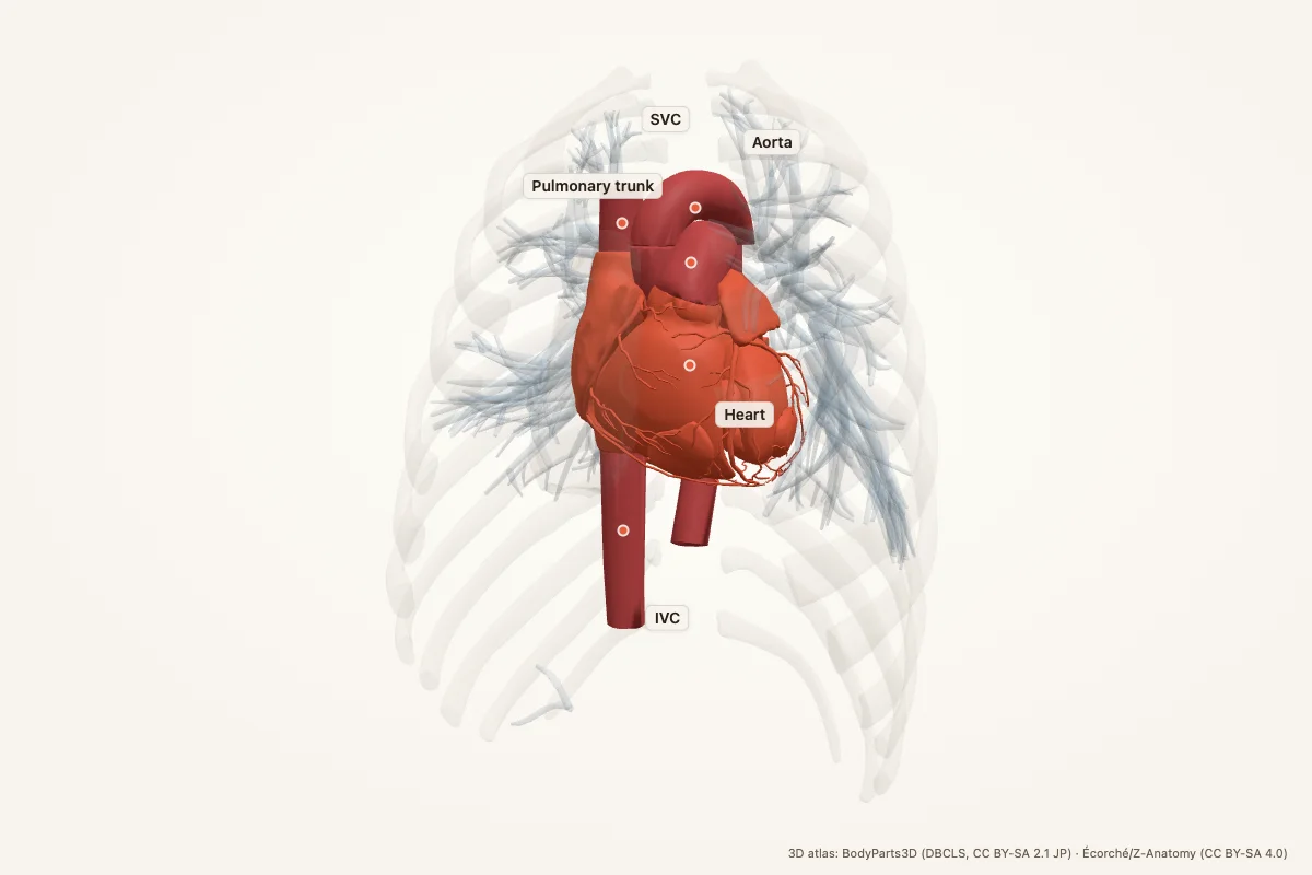
The heart is a four-chambered muscular pump in the mediastinum, behind the sternum and between the lungs. It drives pulmonary and systemic circulation through two atria and two ventricles. Four valves — tricuspid, pulmonary, mitral and aortic — keep blood moving in one direction.
Key facts
| Location | Mediastinum; posterior to the sternum, anterior to the vertebral column, between the lungs |
|---|---|
| Chambers | Right atrium, right ventricle, left atrium, left ventricle |
| Valves | Tricuspid, pulmonary, mitral, aortic |
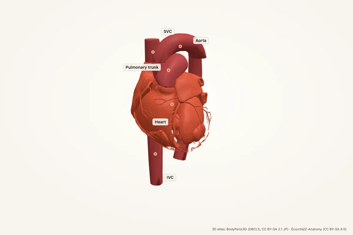
| Blood flow | SVC/IVC → right atrium → right ventricle → pulmonary trunk → lungs → pulmonary veins → left atrium → left ventricle → aorta |
| Coronary supply | Left and right coronary arteries; venous drainage via the coronary sinus |
| Nerve supply | Sympathetic and parasympathetic (vagus) fibres via the cardiac plexus |
| Pericardium | Fibrous and serous pericardium |
| Main function | Pulmonary circulation (right heart) and systemic circulation (left heart) |
Where is the heart located?
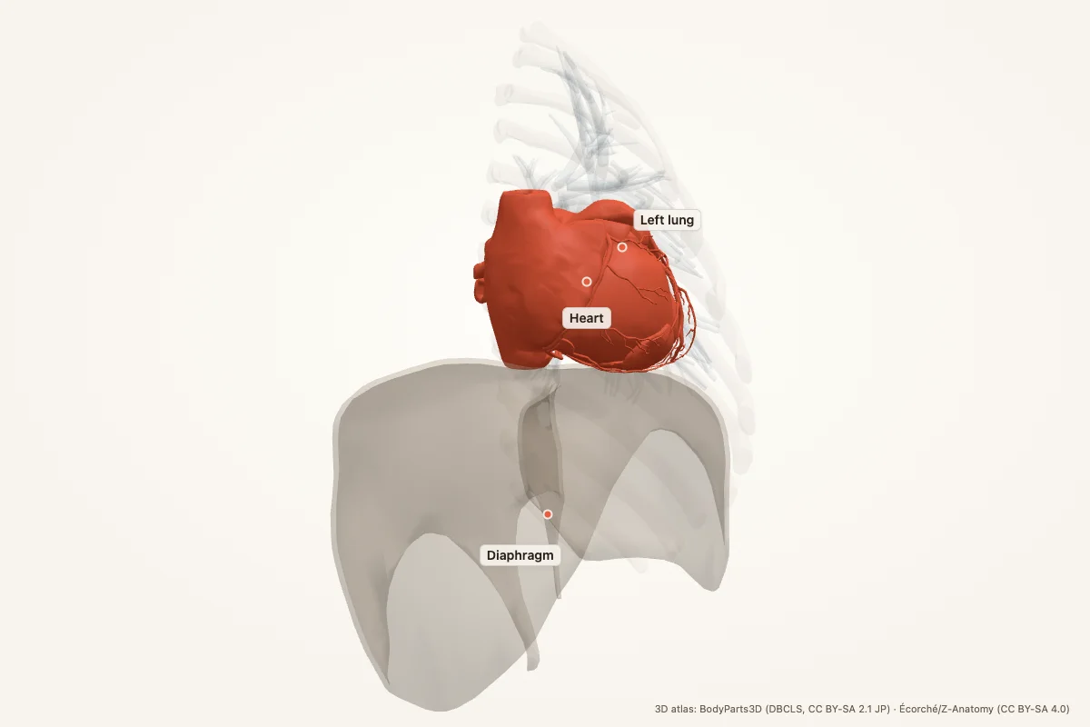
The heart lies in the mediastinum, the central compartment of the thorax. It sits posterior to the sternum and anterior to the vertebral column, between the right and left lungs. About two-thirds of the heart lies left of the midline. The apex points inferiorly, anteriorly and to the left; the base faces superiorly and posteriorly.
What does the heart do?
The right heart receives deoxygenated blood from the body and pumps it to the lungs. The left heart receives oxygenated blood from the lungs and pumps it to the body. The right ventricle drives pulmonary circulation; the left ventricle drives systemic circulation and has a thicker muscular wall.
Chambers and valves
| Chamber | Receives | Pumps to |
|---|---|---|
| Right atrium | Superior vena cava, inferior vena cava, coronary sinus | Right ventricle |
| Right ventricle | Right atrium | Pulmonary trunk |
| Left atrium | Four pulmonary veins | Left ventricle |
| Left ventricle | Left atrium | Aorta |
| Valve | Location | Prevents backflow into |
|---|---|---|
| Tricuspid | Between right atrium and right ventricle | Right atrium |
| Pulmonary | Between right ventricle and pulmonary trunk | Right ventricle |
| Mitral | Between left atrium and left ventricle | Left atrium |
| Aortic | Between left ventricle and aorta | Left ventricle |
Blood flow through the heart
- Deoxygenated blood enters the right atrium through the superior vena cava and inferior vena cava; the coronary sinus also drains into the right atrium.
- Blood passes through the tricuspid valve into the right ventricle.
- The right ventricle contracts and pushes blood through the pulmonary valve into the pulmonary trunk and pulmonary arteries.
- Gas exchange occurs in the lungs; oxygenated blood returns through four pulmonary veins to the left atrium.
- Blood passes through the mitral valve into the left ventricle.
- The left ventricle contracts and pushes blood through the aortic valve into the aorta and systemic circulation.
Coronary blood supply
The left coronary artery arises from the left aortic sinus and divides into the left anterior descending (LAD) and circumflex arteries. The LAD supplies the anterior left ventricle and anterior interventricular septum; the circumflex supplies the left atrium and lateral left ventricle. The right coronary artery supplies the right atrium and right ventricle and usually the SA and AV nodes. Venous blood drains through cardiac veins into the coronary sinus, which opens into the right atrium.
Nerve supply and pericardium
Sympathetic fibres increase heart rate and contractility; parasympathetic fibres from the vagus nerve slow the heart and reduce contractility. The heart is enclosed by the pericardium: a tough fibrous layer and a double-layered serous pericardium (parietal and visceral) with a lubricating pericardial cavity.
Clinical relevance
- Myocardial infarction: coronary occlusion causes myocardial ischaemia and necrosis.
- Angina: reversible myocardial ischaemia, typically on exertion.
- Heart failure: the pump cannot meet the body’s demands, causing congestion and fatigue.
- Valve disease: stenosis or regurgitation alters flow and can overload the chambers.
- Arrhythmias: disorders of impulse generation or conduction disturb the rhythm.
- Pericarditis: inflammation of the pericardium causes chest pain and can lead to effusion.
Mnemonic for the heart valves
“Try Pulling My Aorta” — Tricuspid, Pulmonary, Mitral, Aortic, in blood-flow order.
Pair it with the side rule: right = lungs, left = body. The tricuspid valve is on the right; the mitral valve is on the left.
Heart anatomy quiz
Five questions. Open each answer after you commit to a response.
-
Which chamber pumps blood to the lungs?
Reveal answer
The right ventricle.
-
Which valve sits between the left atrium and the left ventricle?
Reveal answer
The mitral valve.
-
Which vessels carry oxygenated blood from the lungs to the heart?
Reveal answer
The pulmonary veins.
-
Which coronary artery supplies most of the left ventricle?
Reveal answer
The left coronary artery, mainly through the left anterior descending branch.
-
Which ventricular wall is thicker?
Reveal answer
The left ventricular wall, because it pumps against systemic resistance.
Common questions
Where is the heart located?
The heart lies in the mediastinum, posterior to the sternum and anterior to the vertebral column, between the lungs. About two-thirds of its mass sits left of the midline, and its apex points down, forward and to the left.
What are the four chambers of the heart?
The four chambers are the right atrium, right ventricle, left atrium and left ventricle. The right atrium receives deoxygenated blood; the right ventricle pumps it to the lungs. The left atrium receives oxygenated blood; the left ventricle pumps it to the body.
What is the mnemonic for the heart valves?
“Try Pulling My Aorta” gives the four valves in blood-flow order: Tricuspid, Pulmonary, Mitral and Aortic. The tricuspid and mitral valves sit between the atria and ventricles; the pulmonary and aortic valves guard the ventricular outflow tracts.
Which side of the heart pumps blood to the body?
The left side pumps blood to the body. The left ventricle drives systemic circulation through the aorta, so its muscular wall is thicker than the right ventricle’s. The right ventricle pumps only to the lungs.
What artery supplies the heart?
The heart is supplied by the left and right coronary arteries, which arise from the aortic root. The left coronary artery divides into the left anterior descending and circumflex arteries. Venous blood drains through cardiac veins into the coronary sinus and back to the right atrium.
What is the difference between the right and left ventricle?
The right ventricle pumps deoxygenated blood to the lungs through the pulmonary trunk. The left ventricle pumps oxygenated blood to the body through the aorta. The left ventricular wall is thicker because systemic pressure is much higher than pulmonary pressure.
Sources
- Standring, S. (ed.). Gray’s Anatomy, 42nd edition. Elsevier, 2020.
- TeachMeAnatomy — Heart
- Kenhub — Heart
- StatPearls / NCBI Bookshelf — heart anatomy search
3D atlas and image credits
Anatomy models: BodyParts3D, © Database Center for Life Science (DBCLS), CC BY-SA 2.1 Japan. Écorché by Charleeh, derived from BodyParts3D and Z-Anatomy, CC BY-SA 4.0. Rendered in anotemy. Changes: highlighting, framing, labels and background removal. Images: CC BY-SA 4.0.
The 3D models are third-party assets under Creative Commons share-alike licences. They keep those licences; the page text is separate. If you reuse an image, keep the attribution and the share-alike licence.