 anotemy
Sign in
Try it free
Lungs — Anatomy Notes, Diagram & Quiz
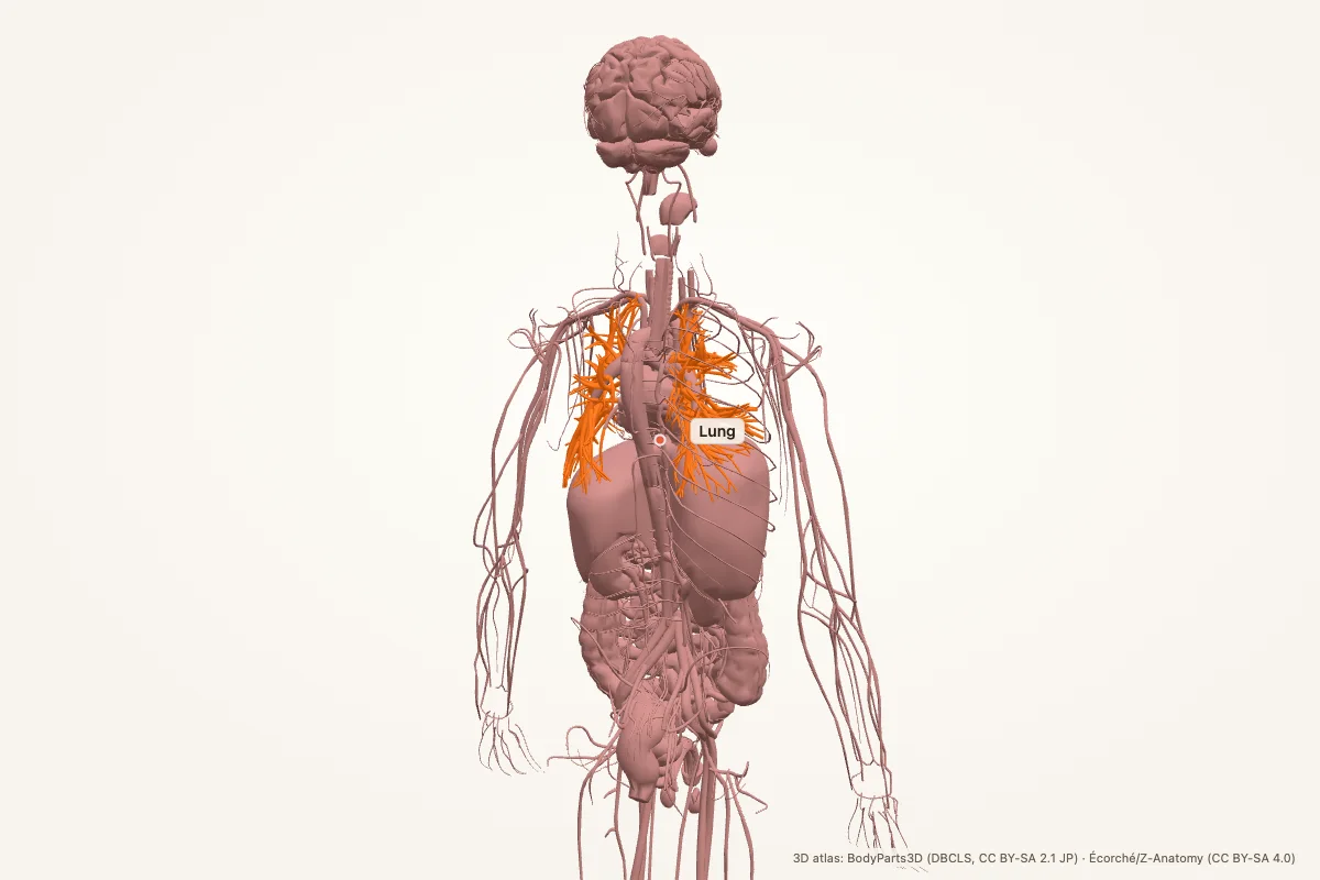
The lungs are paired organs in the thoracic cavity that exchange oxygen and carbon dioxide. The right lung has three lobes; the left has two and a cardiac notch. Air passes through the trachea, bronchi and bronchioles to the alveoli, where gas exchange occurs.
Key facts
| Location | Thoracic cavity, either side of the mediastinum; apex above the clavicle, base on the diaphragm |
|---|---|
| Right lung | Three lobes: upper, middle, lower; oblique and horizontal fissures |
| Left lung | Two lobes: upper and lower; oblique fissure; cardiac notch and lingula |
| Airways | Trachea → main bronchi → lobar bronchi → segmental bronchi → bronchioles → alveoli |
| Pleura | Visceral and parietal pleura with a lubricated pleural cavity |
| Blood supply | Pulmonary arteries for gas exchange; bronchial arteries supply the airway walls |
| Nerve supply | Pulmonary plexus (vagus and sympathetic fibres) |
| Main function | Gas exchange: oxygen into blood, carbon dioxide out |
Where are the lungs located?
The lungs fill the thoracic cavity on either side of the mediastinum. Each lung has an apex that rises above the clavicle and a base that rests on the diaphragm. The right lung is shorter and wider because the liver pushes the right hemidiaphragm upward; the left lung is narrower because the heart occupies the left mediastinum.
The hilum is the root of each lung: the main bronchus, pulmonary artery, pulmonary veins, bronchial vessels, lymphatics and nerves enter or leave there. The lungs are covered by visceral pleura and separated from the chest wall by parietal pleura.
Lobes and fissures
The right lung has three lobes — upper, middle and lower — separated by the oblique and horizontal fissures. The left lung has two lobes, upper and lower, separated by the oblique fissure. The left upper lobe carries the lingula, a tongue-like projection that corresponds to the right middle lobe.
Each lobe is divided into bronchopulmonary segments, each supplied by its own segmental bronchus. The right lung has ten segments and the left has eight to ten, depending on how the segments are counted. This segmental anatomy is why a pneumonia or tumour can be confined to one segment.
Airways and alveoli
The trachea bifurcates at the carina, roughly at the level of T4–T5, into the right and left main bronchi. The right main bronchus is wider, shorter and more vertical, so inhaled foreign bodies are more likely to lodge on the right. The main bronchi divide into lobar, segmental and smaller bronchi, then bronchioles, terminal bronchioles and respiratory bronchioles.
Gas exchange happens in the alveoli, tiny air sacs surrounded by capillaries. The alveolar wall and capillary wall form the blood–air barrier. Type I pneumocytes cover most of the surface; type II pneumocytes secrete surfactant, which lowers surface tension and keeps the alveoli open.
Pleura and pleural cavity
The visceral pleura clings to the lung surface; the parietal pleura lines the thoracic wall, diaphragm and mediastinum. Between them is the pleural cavity, a potential space with a thin film of fluid. Surface tension and negative intrapleural pressure keep the lung expanded against the chest wall.
The pleura reflects at the hilum and forms recesses, including the costodiaphragmatic recess. Fluid collects in these recesses when a patient is upright, which is why a pleural effusion is first seen at the lung base on a chest radiograph.
Blood supply and lymphatic drainage
The pulmonary arteries carry deoxygenated blood from the right ventricle to the lungs, and the pulmonary veins return oxygenated blood to the left atrium. The bronchial arteries, branches of the descending aorta, supply the bronchi, bronchioles and pleura with oxygenated systemic blood.
Lymphatic vessels drain through intrapulmonary and hilar nodes toward the tracheobronchial and mediastinal nodes. This pathway matters in lung cancer staging: tumour spread follows the bronchi and lymphatics toward the hilum and mediastinum.
Muscles of respiration
The diaphragm is the primary muscle of inspiration. It is innervated by the phrenic nerve (C3–C5) and contracts downward, increasing thoracic volume. The external intercostals elevate the ribs. Accessory muscles — scalenes, sternocleidomastoid and pectorals — are recruited when breathing is laboured.
Quiet expiration is mostly passive, driven by elastic recoil. Forced expiration uses the internal intercostals and abdominal muscles. The lungs also have elastic recoil and surfactant to keep breathing efficient.
Clinical relevance
- Pneumonia: infection inflames the alveoli and fills them with exudate, causing consolidation.
- Pneumothorax: air enters the pleural cavity and collapses the lung.
- Asthma: bronchospasm and inflammation narrow the airways, causing wheeze and dyspnoea.
- COPD: emphysema destroys alveolar walls and chronic bronchitis narrows the airways.
- Pulmonary embolism: a clot blocks a pulmonary artery and impairs gas exchange.
- Lung cancer: commonly arises from bronchial epithelium and spreads via lymphatics.
Memory aid: right 3, left 2
Right lung = 3 lobes; left lung = 2 lobes. The heart takes the space on the left, so the left lung gives up the middle lobe and keeps the lingula.
For the airways, follow the branching: trachea → main bronchus → lobar → segmental → bronchiole → alveolus.
Lung anatomy quiz
Five questions. Open each answer after you commit to a response.
-
How many lobes does the right lung have?
Reveal answer
Three: upper, middle and lower.
-
Which lung has the cardiac notch?
Reveal answer
The left lung.
-
Where does the trachea bifurcate?
Reveal answer
At the carina, roughly at the level of T4–T5.
-
Which main bronchus is more likely to receive an inhaled foreign body?
Reveal answer
The right main bronchus, because it is wider, shorter and more vertical.
-
What is the primary muscle of inspiration?
Reveal answer
The diaphragm.
Common questions
Where are the lungs located?
The lungs sit in the thoracic cavity on either side of the mediastinum. Each apex rises above the clavicle and each base rests on the diaphragm. The right lung is shorter because of the liver; the left is narrower because of the heart.
How many lobes does each lung have?
The right lung has three lobes — upper, middle and lower. The left lung has two — upper and lower. The left upper lobe includes the lingula, which corresponds to the right middle lobe.
Why does the left lung have only two lobes?
The heart occupies the left side of the mediastinum, so the left lung has less space. It develops two lobes and a cardiac notch instead of a middle lobe; the lingula is the remnant of that space.
What happens at the alveoli?
Alveoli are the gas-exchange units. Oxygen diffuses across the alveolar wall into the capillary blood, and carbon dioxide diffuses from blood into the alveolar air. Surfactant from type II pneumocytes keeps the alveoli open.
What is the pleura?
The pleura is a double serous membrane. The visceral pleura covers the lung; the parietal pleura lines the chest wall, diaphragm and mediastinum. The pleural cavity between them contains a thin film of fluid that lubricates movement.
What is the carina?
The carina is the ridge where the trachea divides into the right and left main bronchi. It lies roughly at T4–T5 and is a key landmark on bronchoscopy; widening can suggest enlarged mediastinal lymph nodes.
Sources
- Standring, S. (ed.). Gray’s Anatomy, 42nd edition. Elsevier, 2020.
- TeachMeAnatomy — Lungs
- Kenhub — Lungs
- StatPearls / NCBI Bookshelf — lung anatomy search
3D atlas and image credits
Anatomy models: BodyParts3D, © Database Center for Life Science (DBCLS), CC BY-SA 2.1 Japan. Écorché by Charleeh, derived from BodyParts3D and Z-Anatomy, CC BY-SA 4.0. Rendered in anotemy. Changes: highlighting, framing, labels and background removal. Images: CC BY-SA 4.0.
The 3D models are third-party assets under Creative Commons share-alike licences. They keep those licences; the page text is separate. If you reuse an image, keep the attribution and the share-alike licence.