 anotemy
Sign in
Try it free
Femoral artery — Anatomy Notes, Diagram & Quiz
The Femoral artery is located in the thigh. Main arterial supply to lower limb. The Femoral artery is a continuation of the external iliac artery distal to the inguinal ligament.
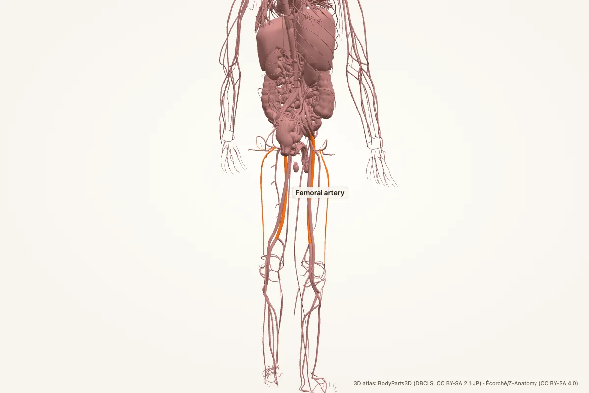
Key facts
| Location | Thigh |
|---|---|
| Main function | Main arterial supply to lower limb. |
| Layer | organs |
| Key fact 1 | The Femoral artery is a continuation of the external iliac artery distal to the inguinal ligament. |
| Key fact 2 | The Femoral artery supplies blood to the thigh and continues as the popliteal artery behind the knee. |
| Key fact 3 | The Femoral artery is palpable in the femoral triangle and is used for arterial access in catheterization. |
Where is the Femoral artery located?
The Femoral artery is located in the thigh.
The Femoral artery is a continuation of the external iliac artery distal to the inguinal ligament.
What does the Femoral artery do?
Main arterial supply to lower limb.
The Femoral artery supplies blood to the thigh and continues as the popliteal artery behind the knee.
What to know for exams
- The Femoral artery is a continuation of the external iliac artery distal to the inguinal ligament.
- The Femoral artery supplies blood to the thigh and continues as the popliteal artery behind the knee.
- The Femoral artery is palpable in the femoral triangle and is used for arterial access in catheterization.
It belongs to the organs layer. Learn its location, relations, and the structures it works with.
Clinical relevance
The Femoral artery is palpable in the femoral triangle and is used for arterial access in catheterization.
Link the femoral artery to its blood supply, innervation and relations. That is how exam questions usually test it.
Memory aid: Femoral artery
The Femoral artery is a continuation of the external iliac artery distal to the inguinal ligament. The Femoral artery supplies blood to the thigh and continues as the popliteal artery behind the knee. The Femoral artery is palpable in the femoral triangle and is used for arterial access in catheterization.
Chain it as location → function → clinical link when you revise.
Femoral artery anatomy quiz
Five questions. Open each answer after you commit to a response.
-
Where is the Femoral artery located?
Reveal answer
Thigh
-
What is the main function of the Femoral artery?
Reveal answer
Main arterial supply to lower limb.
-
Give one key fact about the Femoral artery.
Reveal answer
The Femoral artery is a continuation of the external iliac artery distal to the inguinal ligament.
-
Give another key fact about the Femoral artery.
Reveal answer
The Femoral artery supplies blood to the thigh and continues as the popliteal artery behind the knee.
-
What clinical link is associated with the Femoral artery?
Reveal answer
The Femoral artery is palpable in the femoral triangle and is used for arterial access in catheterization.
Common questions
Where is the Femoral artery located?
The Femoral artery is located in the thigh. The Femoral artery is a continuation of the external iliac artery distal to the inguinal ligament.
What does the Femoral artery do?
Main arterial supply to lower limb. The Femoral artery supplies blood to the thigh and continues as the popliteal artery behind the knee.
What is a key fact about the Femoral artery?
The Femoral artery is palpable in the femoral triangle and is used for arterial access in catheterization.
What layer does the Femoral artery belong to?
It belongs to the organs layer of the body.
What should I remember for exams?
The Femoral artery is a continuation of the external iliac artery distal to the inguinal ligament. The Femoral artery supplies blood to the thigh and continues as the popliteal artery behind the knee. The Femoral artery is palpable in the femoral triangle and is used for arterial access in catheterization.
Sources
- Standring, S. (ed.). Gray’s Anatomy, 42nd edition. Elsevier, 2020.
- TeachMeAnatomy — anatomy library
- Kenhub — anatomy library
- StatPearls / NCBI Bookshelf — Femoral artery search
3D atlas and image credits
Anatomy models: BodyParts3D, © Database Center for Life Science (DBCLS), CC BY-SA 2.1 Japan. Écorché by Charleeh, derived from BodyParts3D and Z-Anatomy, CC BY-SA 4.0. Rendered in anotemy. Changes: highlighting, framing, labels and background removal. Images: CC BY-SA 4.0.
The 3D models are third-party assets under Creative Commons share-alike licences. They keep those licences; the page text is separate. If you reuse an image, keep the attribution and the share-alike licence.