 anotemy
Sign in
Try it free
Scapula — Anatomy Notes, Diagram & Quiz
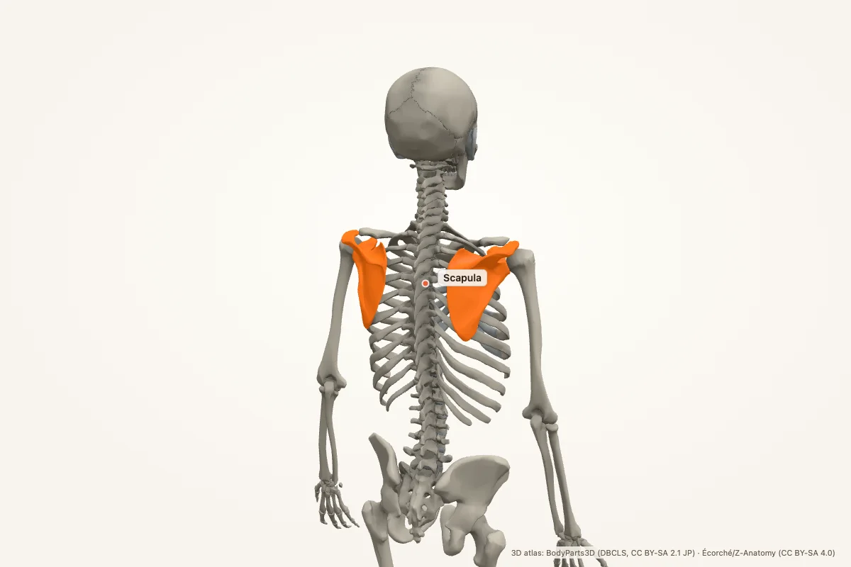
The scapula is the shoulder blade, a flat triangular bone on the posterior chest wall. It connects the clavicle to the humerus and provides attachment for the rotator cuff and many shoulder muscles. Key landmarks include the spine, acromion, coracoid process and glenoid cavity.
Key facts
| Region | Posterior shoulder / upper back |
|---|---|
| Type | Flat triangular bone |
| Borders and angles | Superior, medial and lateral borders; superior, inferior and lateral angles |
| Key landmarks | Spine, acromion, coracoid process, glenoid cavity, suprascapular notch |
| Fossae | Supraspinous, infraspinous and subscapular fossae |
| Articulations | Acromioclavicular joint and glenohumeral joint |
| Main muscles | Rotator cuff, deltoid, trapezius, rhomboids, serratus anterior, levator scapulae |
| Clinical | Winged scapula, scapular fracture, shoulder dislocation, suprascapular nerve entrapment |
Where is the scapula located?
The scapula lies on the posterior chest wall, over the ribs, from the second to the seventh rib. It is a flat triangular bone with a medial border near the spine, a lateral border toward the axilla, and a superior border at the base of the neck.
The scapula is highly mobile: it glides and rotates on the chest wall during arm elevation. It connects to the clavicle at the acromioclavicular joint and to the humerus at the glenohumeral joint.
Landmarks
The spine of the scapula runs across the posterior surface and ends laterally as the acromion, which forms the point of the shoulder. The coracoid process projects anteriorly below the clavicle and gives attachment to the short head of biceps, coracobrachialis and pectoralis minor.
The glenoid cavity is the shallow socket for the humeral head. The suprascapular notch is on the superior border and transmits the suprascapular nerve. The posterior surface has supraspinous and infraspinous fossae; the anterior surface has the subscapular fossa.
Muscle attachments
The rotator cuff attaches around the glenoid: supraspinatus in the supraspinous fossa, infraspinatus in the infraspinous fossa, teres minor on the lateral border, and subscapularis in the subscapular fossa. These muscles stabilise the humeral head.
The trapezius and rhomboids attach along the medial border; the serratus anterior attaches along the medial border anteriorly; the levator scapulae attaches at the superior angle; and the deltoid attaches to the spine and acromion. The long head of triceps attaches to the infraglenoid tubercle.
Clinical relevance
- Winged scapula: serratus anterior weakness (long thoracic nerve) makes the medial border lift off the chest wall.
- Scapular fracture: usually from high-energy trauma; associated chest and shoulder injuries are common.
- Shoulder dislocation: the humeral head leaves the glenoid, often anteriorly.
- Suprascapular nerve entrapment: causes posterior shoulder pain and weakness of supraspinatus and infraspinatus.
- Acromioclavicular joint injury: a shoulder separation after a fall onto the shoulder.
- Scapular dyskinesis: abnormal scapular motion contributes to shoulder impingement.
Mnemonic: SITS around the scapula
SITS — Supraspinatus, Infraspinatus, Teres minor, Subscapularis. These are the rotator cuff muscles that attach to the scapula and humerus.
Landmark order on the posterior scapula: spine → acromion laterally, coracoid anteriorly, glenoid laterally.
Scapula anatomy quiz
Five questions. Open each answer after you commit to a response.
-
Which bony projection forms the point of the shoulder?
Reveal answer
The acromion.
-
Which process projects anteriorly below the clavicle?
Reveal answer
The coracoid process.
-
Which cavity receives the head of the humerus?
Reveal answer
The glenoid cavity.
-
Which nerve passes through the suprascapular notch?
Reveal answer
The suprascapular nerve.
-
Which muscle weakness causes a winged scapula?
Reveal answer
Serratus anterior, from long thoracic nerve injury.
Common questions
Where is the scapula located?
The scapula is on the posterior chest wall, over the second to seventh ribs. It is a flat triangular bone that connects the clavicle to the humerus and moves over the thorax during arm elevation.
What are the main landmarks of the scapula?
The spine, acromion, coracoid process, glenoid cavity, suprascapular notch, and the supraspinous, infraspinous and subscapular fossae. The medial, lateral and superior borders and the superior, inferior and lateral angles are also key.
Which muscles attach to the scapula?
The rotator cuff (supraspinatus, infraspinatus, teres minor, subscapularis), deltoid, trapezius, rhomboids, serratus anterior, levator scapulae, biceps short head, coracobrachialis, pectoralis minor and triceps long head all attach to the scapula.
What causes a winged scapula?
A winged scapula is usually caused by weakness of serratus anterior after injury to the long thoracic nerve. The medial border of the scapula lifts away from the chest wall, especially when pushing forward against a wall.
What is the glenoid cavity?
The glenoid cavity is the shallow socket on the lateral scapula that articulates with the humeral head. The glenoid labrum deepens the socket, and the rotator cuff stabilises the joint.
Is the scapula part of the axial or appendicular skeleton?
The scapula is part of the appendicular skeleton. It connects the upper limb to the axial skeleton through the clavicle and sternum, and it moves with the arm rather than forming part of the spine or rib cage.
Sources
- Standring, S. (ed.). Gray’s Anatomy, 42nd edition. Elsevier, 2020.
- TeachMeAnatomy — Scapula
- Kenhub — Scapula
- StatPearls / NCBI Bookshelf — scapula anatomy search
3D atlas and image credits
Anatomy models: BodyParts3D, © Database Center for Life Science (DBCLS), CC BY-SA 2.1 Japan. Écorché by Charleeh, derived from BodyParts3D and Z-Anatomy, CC BY-SA 4.0. Rendered in anotemy. Changes: highlighting, framing, labels and background removal. Images: CC BY-SA 4.0.
The 3D models are third-party assets under Creative Commons share-alike licences. They keep those licences; the page text is separate. If you reuse an image, keep the attribution and the share-alike licence.