 anotemy
Sign in
Try it free
Humerus — Anatomy Notes, Diagram & Quiz
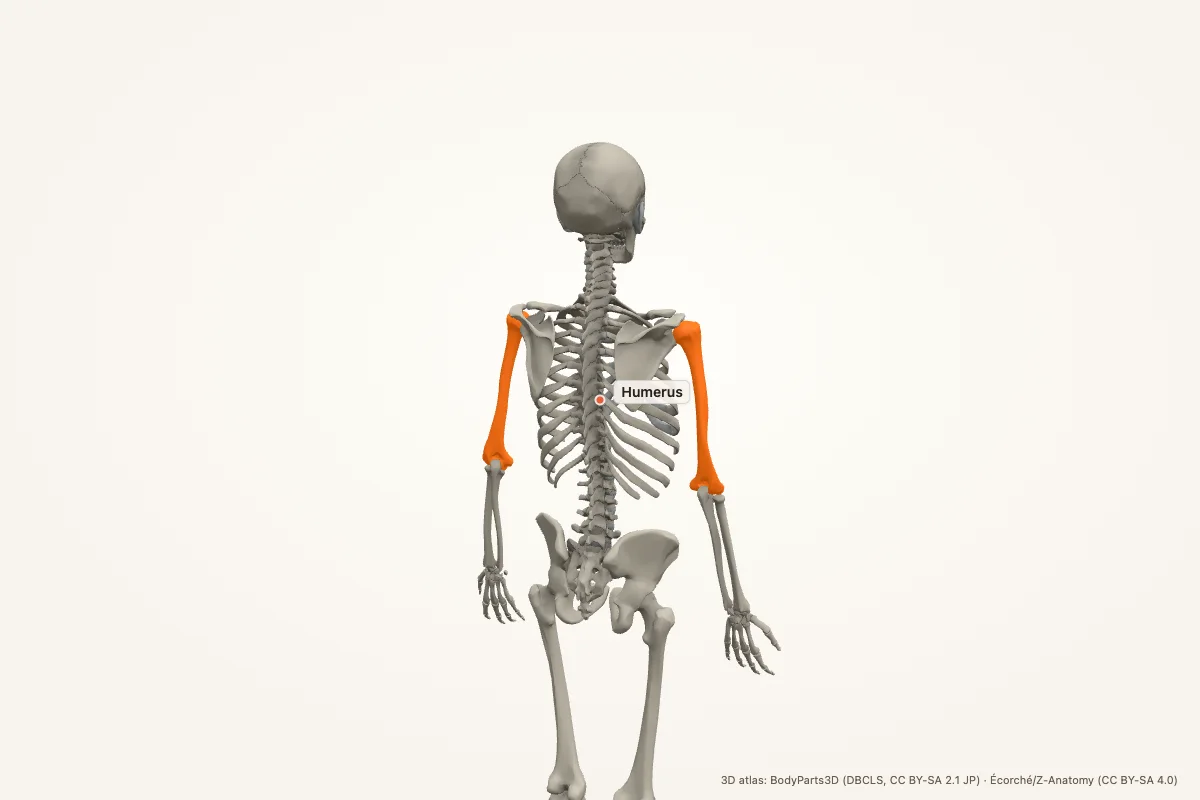
The humerus is the bone of the arm, running from the shoulder to the elbow. Its proximal head articulates with the glenoid; its distal condyles articulate with the radius and ulna. Key landmarks include the surgical neck, radial groove and medial epicondyle.
Key facts
| Region | Arm; shoulder to elbow |
|---|---|
| Proximal landmarks | Head, anatomical neck, surgical neck, greater and lesser tubercles, intertubercular groove |
| Shaft | Deltoid tuberosity, radial groove |
| Distal landmarks | Capitulum, trochlea, medial and lateral epicondyles, olecranon fossa |
| Articulations | Glenohumeral joint, elbow joint |
| Nerves at risk | Axillary at the surgical neck, radial in the radial groove, ulnar behind the medial epicondyle |
| Main muscles | Deltoid, pectoralis major, latissimus dorsi, biceps, triceps, brachialis |
| Clinical | Proximal humerus fracture, shaft fracture, supracondylar fracture, radial nerve palsy |
Where is the humerus located?
The humerus is the single bone of the arm, running from the shoulder joint to the elbow joint. Its proximal end forms the head that fits into the glenoid cavity of the scapula. Its distal end expands into the condyles that articulate with the radius and ulna.
The humerus is surrounded by the deltoid, pectoralis major, latissimus dorsi, biceps, triceps and brachialis. The radial nerve runs in a groove on its posterior surface, and the ulnar nerve passes behind the medial epicondyle.
Proximal humerus
The head is rounded and faces medially, posteriorly and slightly superiorly. The anatomical neck is the old epiphyseal line; the surgical neck is the narrower region below the tubercles and is a common fracture site. The axillary nerve winds around the surgical neck.
The greater tubercle is lateral and receives the supraspinatus, infraspinatus and teres minor. The lesser tubercle is anterior and receives the subscapularis. The intertubercular groove runs between them and carries the long head of biceps.
Humeral shaft
The shaft is roughly cylindrical. The deltoid tuberosity is a rough area on the lateral surface where the deltoid inserts. The radial groove runs obliquely down the posterior surface and carries the radial nerve and profunda brachii vessels.
The shaft gives attachment to the brachialis anteriorly, the triceps posteriorly, and the brachioradialis and extensor muscles distally. Fractures at mid-shaft can injure the radial nerve and cause wrist drop.
Distal humerus
The distal humerus expands into the capitulum laterally and the trochlea medially. The capitulum articulates with the radius; the trochlea articulates with the ulna. The olecranon fossa is a deep posterior depression that receives the olecranon during extension.
The medial and lateral epicondyles are palpable. The ulnar nerve passes behind the medial epicondyle, which is why hitting the “funny bone” causes tingling in the little and ring fingers. The common flexor origin is on the medial epicondyle; the common extensor origin is on the lateral epicondyle.
Clinical relevance
- Proximal humerus fracture: common in older adults; the axillary nerve is at risk.
- Mid-shaft fracture: the radial nerve can be injured, causing wrist drop.
- Supracondylar fracture: common in children; the brachial artery and median nerve are at risk.
- Ulnar neuropathy: compression behind the medial epicondyle causes numbness and weakness in the hand.
- Shoulder dislocation: the humeral head can displace anteriorly or posteriorly; axillary nerve function must be checked.
- Epicondylitis: tendinopathy at the common flexor or extensor origin causes medial or lateral elbow pain.
Memory aid: nerves around the humerus
Axillary at the surgical neck; radial in the radial groove; ulnar behind the medial epicondyle.
For the distal articulations: capitulum = radius; trochlea = ulna. The lateral epicondyle is the common extensor origin; the medial epicondyle is the common flexor origin.
Humerus anatomy quiz
Five questions. Open each answer after you commit to a response.
-
Which nerve is at risk at the surgical neck of the humerus?
Reveal answer
The axillary nerve.
-
Which nerve runs in the radial groove?
Reveal answer
The radial nerve.
-
Which distal humeral surface articulates with the radius?
Reveal answer
The capitulum.
-
Which nerve passes behind the medial epicondyle?
Reveal answer
The ulnar nerve.
-
Which muscle inserts on the deltoid tuberosity?
Reveal answer
The deltoid.
Common questions
Where is the humerus located?
The humerus is the bone of the arm, between the shoulder and elbow. Its head articulates with the glenoid cavity of the scapula, and its distal condyles articulate with the radius and ulna. It is the only bone in the arm.
What is the surgical neck of the humerus?
The surgical neck is the narrowed region below the greater and lesser tubercles. It is a common fracture site, and the axillary nerve winds around it. A fracture here can cause deltoid weakness and numbness over the shoulder.
Why does a mid-shaft humeral fracture cause wrist drop?
The radial nerve runs in the radial groove on the posterior humeral shaft. A fracture there can injure the nerve, paralysing the wrist and finger extensors. The result is wrist drop and sensory loss over the dorsum of the hand.
Which muscles attach to the humerus?
The rotator cuff attaches to the tubercles; deltoid to the deltoid tuberosity; pectoralis major, latissimus dorsi and teres major to the intertubercular groove; brachialis to the anterior shaft; triceps to the posterior shaft; and forearm muscles to the epicondyles.
What is the difference between the anatomical and surgical neck?
The anatomical neck is the old epiphyseal plate around the head. The surgical neck is the narrower region below the tubercles. The surgical neck is more often fractured and is closely related to the axillary nerve.
Which epicondyle is the common extensor origin?
The lateral epicondyle is the common extensor origin, and tendinopathy there is lateral epicondylitis (tennis elbow). The medial epicondyle is the common flexor origin, and tendinopathy there is medial epicondylitis (golfer’s elbow).
Sources
- Standring, S. (ed.). Gray’s Anatomy, 42nd edition. Elsevier, 2020.
- TeachMeAnatomy — Humerus
- Kenhub — Humerus
- StatPearls / NCBI Bookshelf — humerus anatomy search
3D atlas and image credits
Anatomy models: BodyParts3D, © Database Center for Life Science (DBCLS), CC BY-SA 2.1 Japan. Écorché by Charleeh, derived from BodyParts3D and Z-Anatomy, CC BY-SA 4.0. Rendered in anotemy. Changes: highlighting, framing, labels and background removal. Images: CC BY-SA 4.0.
The 3D models are third-party assets under Creative Commons share-alike licences. They keep those licences; the page text is separate. If you reuse an image, keep the attribution and the share-alike licence.