 anotemy
Sign in
Try it free
Kidneys — Anatomy Notes, Diagram & Quiz
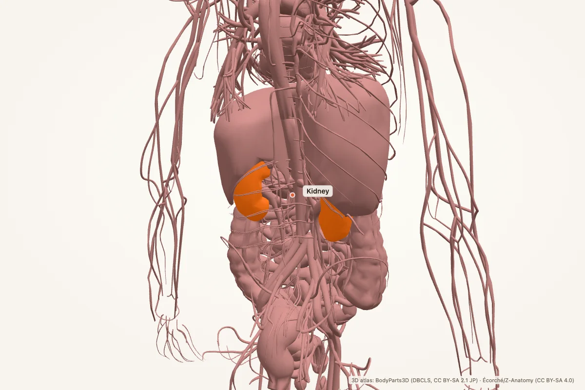
The kidneys are paired retroperitoneal organs that filter blood, remove waste and regulate fluid, electrolyte and acid-base balance. Each kidney has a cortex, medulla and renal pelvis; the nephron is its functional unit. The right kidney usually sits slightly lower than the left because of the liver.
Key facts
| Location | Retroperitoneal, on the posterior abdominal wall, roughly T12–L3 |
|---|---|
| Right vs left | The right kidney usually sits slightly lower because the liver occupies the right upper abdomen |
| Layers | Fibrous capsule, cortex, medulla (renal pyramids), renal pelvis, ureter |
| Functional unit | Nephron: glomerulus, Bowman’s capsule, proximal tubule, loop of Henle, distal tubule, collecting duct |
| Blood supply | Renal artery → segmental → interlobar → arcuate → interlobular arteries |
| Venous drainage | Renal vein → inferior vena cava |
| Hormones | Renin, erythropoietin, calcitriol (active vitamin D) |
| Main function | Filtration, reabsorption, secretion and excretion; fluid, electrolyte and acid-base balance |
Where are the kidneys located?
The kidneys lie retroperitoneally on the posterior abdominal wall, one on each side of the vertebral column, roughly between T12 and L3. They are surrounded by perirenal fat and enclosed by the renal fascia. The adrenal glands sit on top of each kidney.
The right kidney is usually slightly lower than the left because the liver occupies the right upper quadrant. Each kidney has a medial hilum where the renal artery, renal vein and ureter enter or leave. The upper poles lie nearer the midline than the lower poles.
External and internal structure
A fibrous capsule surrounds each kidney. Inside, the cortex is the outer layer containing glomeruli and convoluted tubules. The medulla contains renal pyramids, whose apices (renal papillae) project into minor calyces. Minor calyces join to form major calyces, which drain into the renal pelvis and then the ureter.
The kidney has a concave medial border with the hilum and a convex lateral border. The renal sinus contains the pelvis, calyces, vessels and fat. The cortex also contains renal columns that extend between the pyramids.
The nephron and urine formation
The nephron is the functional unit of the kidney, and each kidney has about one million. Blood enters the glomerulus through the afferent arteriole and leaves through the efferent arteriole. Filtration occurs across the glomerular filtration barrier into Bowman’s capsule.
The proximal convoluted tubule reabsorbs most filtered water, sodium, glucose and amino acids. The loop of Henle creates a medullary osmotic gradient; the distal convoluted tubule and collecting duct fine-tune sodium, potassium, water and acid-base balance under hormonal control.
Blood supply and drainage
Each kidney receives a renal artery from the abdominal aorta. The artery divides into segmental arteries, then interlobar, arcuate and interlobular arteries. Afferent arterioles feed the glomeruli; efferent arterioles form the peritubular capillaries and vasa recta that surround the tubules.
Venous blood drains through interlobular, arcuate and interlobar veins into the renal vein, which drains into the inferior vena cava. The kidneys also have rich lymphatic drainage toward the para-aortic nodes.
Hormonal and metabolic roles
The kidneys produce renin, which activates the renin–angiotensin– aldosterone system and raises blood pressure. They produce erythropoietin, which stimulates red blood cell production, and calcitriol, the active form of vitamin D, which supports calcium absorption and bone health.
They also regulate potassium, sodium, bicarbonate and pH, and they excrete metabolic waste such as urea and creatinine. This is why kidney failure affects blood pressure, bone health and red cell production, not just urine output.
Clinical relevance
- Acute kidney injury: a rapid fall in filtration, often from dehydration, sepsis or nephrotoxins.
- Chronic kidney disease: progressive loss of function, commonly from diabetes and hypertension.
- Kidney stones: crystals form in the collecting system and cause colicky flank pain.
- Pyelonephritis: infection of the renal pelvis and parenchyma, usually ascending from the bladder.
- Renal artery stenosis: narrowed renal artery activates renin and causes secondary hypertension.
- Polycystic kidney disease: inherited cysts enlarge the kidneys and reduce function.
Memory aid: right lower, left higher
The right kidney sits lower because the liver takes the space above it. Both kidneys are retroperitoneal — behind the peritoneum, against the posterior abdominal wall.
For the drainage path: papilla → minor calyx → major calyx → renal pelvis → ureter → bladder.
Kidney anatomy quiz
Five questions. Open each answer after you commit to a response.
-
Where do the kidneys lie?
Reveal answer
Retroperitoneally on the posterior abdominal wall, roughly T12–L3.
-
Which kidney is usually lower?
Reveal answer
The right kidney, because the liver occupies the space above it.
-
What is the functional unit of the kidney?
Reveal answer
The nephron.
-
Which artery supplies each kidney?
Reveal answer
The renal artery, a branch of the abdominal aorta.
-
Which hormone produced by the kidneys stimulates red blood cell production?
Reveal answer
Erythropoietin.
Common questions
Where are the kidneys located?
The kidneys are retroperitoneal organs on the posterior abdominal wall, roughly from T12 to L3. The right kidney is usually slightly lower than the left because the liver occupies the right upper abdomen. Each kidney has an adrenal gland above it.
What do the kidneys do?
They filter blood, remove metabolic waste, and regulate water, electrolytes, acid-base balance and blood pressure. They also produce renin, erythropoietin and active vitamin D, and they help control calcium and phosphate balance.
Why is the right kidney lower than the left?
The liver occupies the right upper quadrant and pushes the right kidney downward. The left kidney has more room under the diaphragm and sits slightly higher. This is a normal positional difference, not a disease.
What is a nephron?
A nephron is the functional unit of the kidney. It consists of a glomerulus, Bowman’s capsule, proximal tubule, loop of Henle, distal tubule and collecting duct. Each kidney contains about one million nephrons.
What artery supplies the kidney?
The renal artery arises from the abdominal aorta and divides into segmental, interlobar, arcuate and interlobular arteries. Blood then passes through afferent arterioles into the glomeruli and drains through the renal vein to the inferior vena cava.
What is the renal pelvis?
The renal pelvis is the funnel-shaped collecting structure at the hilum. Minor calyces drain into major calyces, which join to form the pelvis; the pelvis continues as the ureter. Urine flows from the papillae into the calyces and out through the pelvis.
Sources
- Standring, S. (ed.). Gray’s Anatomy, 42nd edition. Elsevier, 2020.
- TeachMeAnatomy — Kidneys
- Kenhub — Kidneys
- StatPearls / NCBI Bookshelf — kidney anatomy search
3D atlas and image credits
Anatomy models: BodyParts3D, © Database Center for Life Science (DBCLS), CC BY-SA 2.1 Japan. Écorché by Charleeh, derived from BodyParts3D and Z-Anatomy, CC BY-SA 4.0. Rendered in anotemy. Changes: highlighting, framing, labels and background removal. Images: CC BY-SA 4.0.
The 3D models are third-party assets under Creative Commons share-alike licences. They keep those licences; the page text is separate. If you reuse an image, keep the attribution and the share-alike licence.