 anotemy
Sign in
Try it free
Stomach — Anatomy Notes, Diagram & Quiz
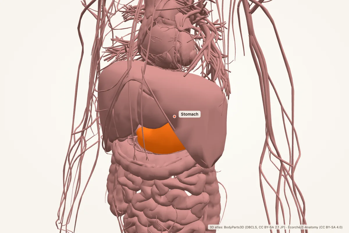
The stomach is a muscular organ in the left upper quadrant between the esophagus and duodenum. It stores and mixes food, secretes acid and pepsinogen, and delivers chyme to the small intestine. Its wall has mucosa, submucosa, muscularis externa and serosa.
Key facts
| Location | Left upper quadrant, between the esophagus and duodenum, under the diaphragm |
|---|---|
| Parts | Cardia, fundus, body, pylorus (antrum and pyloric canal) |
| Curvatures | Greater curvature and lesser curvature |
| Wall layers | Mucosa, submucosa, muscularis externa (oblique, circular, longitudinal), serosa |
| Glands and cells | Parietal cells (HCl, intrinsic factor), chief cells (pepsinogen), G cells (gastrin), mucous cells |
| Sphincters | Lower esophageal sphincter and pyloric sphincter |
| Blood supply | Left gastric, right gastric, short gastric and gastroepiploic arteries |
| Nerve supply | Vagus nerve (parasympathetic) and celiac plexus (sympathetic) |
Where is the stomach located?
The stomach lies in the left upper quadrant, between the esophagus and the duodenum. It sits under the diaphragm, anterior to the pancreas and posterior to the liver, which overlaps it anteriorly. The stomach is intraperitoneal and is connected to surrounding organs by the greater and lesser omenta.
The cardia is the region where the esophagus enters. The fundus is the dome above the cardia. The body is the main reservoir, and the pylorus is the distal outlet. The pyloric sphincter controls emptying into the duodenum.
Parts and curvatures
The stomach has four parts: cardia, fundus, body and pylorus. The pylorus includes the antrum and the pyloric canal. The greater curvature runs along the convex lower border; the lesser curvature runs along the concave upper border.
The greater omentum hangs from the greater curvature, and the lesser omentum connects the lesser curvature to the liver. These peritoneal attachments carry blood vessels, lymphatics and nerves, and they help define the surgical planes around the stomach.
Wall and gastric glands
The stomach wall has four layers: mucosa, submucosa, muscularis externa and serosa. The mucosa is folded into rugae when the stomach is empty and contains gastric pits and glands. The muscularis externa has an extra oblique layer in addition to the circular and longitudinal layers.
Parietal cells secrete hydrochloric acid and intrinsic factor. Chief cells secrete pepsinogen, which becomes pepsin in acid. G cells secrete gastrin, which stimulates acid production. Mucous cells and bicarbonate protect the lining from acid and pepsin.
Secretion and digestion
The stomach stores food, mixes it with gastric juice and gradually empties chyme into the duodenum. Acid denatures proteins and activates pepsin; pepsin begins protein digestion. Intrinsic factor is essential for vitamin B12 absorption in the terminal ileum.
Gastric secretion is controlled by neural and hormonal signals: acetylcholine from the vagus, gastrin from G cells and histamine from enterochromaffin-like cells. Somatostatin and prostaglandins inhibit acid secretion. The stomach absorbs little water and few nutrients; its main job is preparation, not absorption.
Blood supply and nerve supply
The stomach receives blood from the celiac trunk. The left gastric artery supplies the lesser curvature, the right gastric artery joins it, and the short gastric and gastroepiploic arteries supply the greater curvature. Venous blood drains into the portal system.
The vagus nerve provides parasympathetic supply and stimulates acid secretion and motility. Sympathetic fibres from the celiac plexus reduce motility and constrict vessels. Lymphatic drainage follows the vessels toward the celiac nodes.
Clinical relevance
- Peptic ulcer: acid and pepsin damage the mucosa, often with H. pylori or NSAID use.
- Gastritis: inflammation of the gastric mucosa, which can be acute or chronic.
- GERD: reflux of gastric acid into the esophagus causes heartburn and mucosal injury.
- Gastric cancer: often associated with chronic H. pylori infection and atrophic gastritis.
- Pernicious anemia: loss of intrinsic factor prevents vitamin B12 absorption.
- Dumping syndrome: rapid gastric emptying after surgery causes cramps, diarrhoea and flushing.
Memory aid: CFBP and parietal cells
CFBP — Cardia, Fundus, Body, Pylorus. That is the stomach from proximal to distal.
Parietal cells make acid + intrinsic factor. Intrinsic factor is the one people forget; without it, vitamin B12 absorption fails in the terminal ileum.
Stomach anatomy quiz
Five questions. Open each answer after you commit to a response.
-
What are the four parts of the stomach?
Reveal answer
Cardia, fundus, body and pylorus.
-
Which cells secrete hydrochloric acid and intrinsic factor?
Reveal answer
Parietal cells.
-
Which cells secrete pepsinogen?
Reveal answer
Chief cells.
-
Which nerve provides parasympathetic supply to the stomach?
Reveal answer
The vagus nerve.
-
Which vitamin requires intrinsic factor for absorption?
Reveal answer
Vitamin B12.
Common questions
Where is the stomach located?
The stomach sits in the left upper quadrant, under the diaphragm and between the esophagus and duodenum. The liver lies anterior to it. It is intraperitoneal and attached by the greater and lesser omenta.
What are the parts of the stomach?
The four parts are the cardia, fundus, body and pylorus. The cardia receives the esophagus, the fundus is the dome above it, the body is the main reservoir, and the pylorus controls emptying into the duodenum.
Why doesn’t stomach acid digest the stomach?
Mucous cells secrete a protective mucus and bicarbonate layer that keeps acid away from the epithelium. Prostaglandins support mucosal blood flow and mucus production. When this barrier fails, peptic ulcers can develop.
What is intrinsic factor?
Intrinsic factor is a protein secreted by parietal cells. It binds vitamin B12 in the small intestine and is required for absorption in the terminal ileum. Loss of intrinsic factor causes pernicious anaemia.
What is H. pylori?
Helicobacter pylori is a bacterium that colonises the gastric mucosa. It causes chronic gastritis and is strongly associated with peptic ulcers and gastric cancer. Treatment usually combines acid suppression with antibiotics.
What does the stomach absorb?
The stomach absorbs little water and few nutrients. It absorbs some alcohol, aspirin and certain drugs, but its main roles are storage, mixing and chemical digestion. Most absorption happens in the small intestine.
Sources
- Standring, S. (ed.). Gray’s Anatomy, 42nd edition. Elsevier, 2020.
- TeachMeAnatomy — Stomach
- Kenhub — Stomach
- StatPearls / NCBI Bookshelf — stomach anatomy search
3D atlas and image credits
Anatomy models: BodyParts3D, © Database Center for Life Science (DBCLS), CC BY-SA 2.1 Japan. Écorché by Charleeh, derived from BodyParts3D and Z-Anatomy, CC BY-SA 4.0. Rendered in anotemy. Changes: highlighting, framing, labels and background removal. Images: CC BY-SA 4.0.
The 3D models are third-party assets under Creative Commons share-alike licences. They keep those licences; the page text is separate. If you reuse an image, keep the attribution and the share-alike licence.