 anotemy
Sign in
Try it free
Liver — Anatomy Notes, Diagram & Quiz
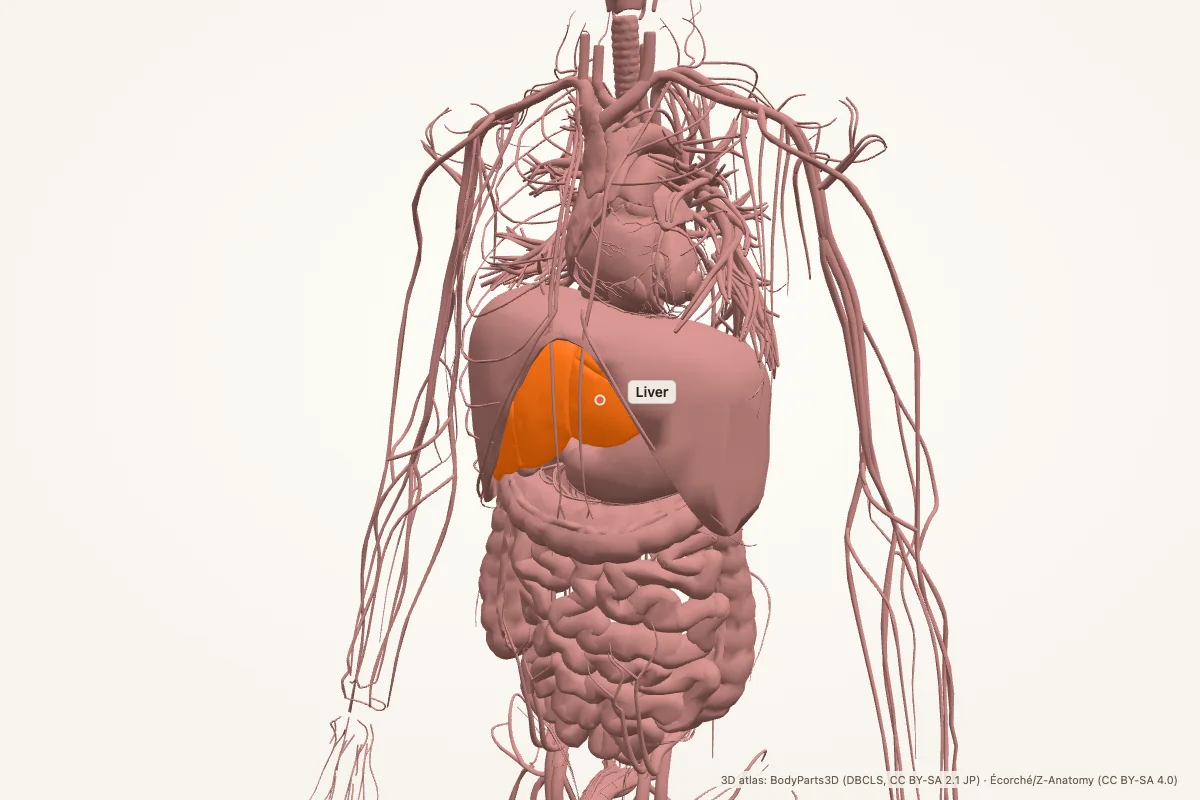
The liver is the largest internal organ, sitting in the right upper quadrant under the diaphragm. It performs metabolism, detoxification, protein synthesis and bile production. It receives blood from the portal vein and hepatic artery and drains bile through the biliary tree.
Key facts
| Location | Right upper quadrant, under the diaphragm, protected by ribs 7–11 |
|---|---|
| Lobes | Right, left, caudate and quadrate |
| Segments | Couinaud segments I–VIII, based on portal and hepatic vein branches |
| Blood supply | Portal vein (~75%) and hepatic artery (~25%) |
| Venous drainage | Hepatic veins → inferior vena cava |
| Bile drainage | Hepatic ducts → common hepatic duct → cystic duct → common bile duct → duodenum |
| Main functions | Metabolism, detoxification, protein synthesis, bile production, storage |
| Key cells | Hepatocytes, Kupffer cells, cholangiocytes, hepatic stellate cells |
Where is the liver located?
The liver occupies the right upper quadrant of the abdomen, tucked under the right hemidiaphragm. It extends across the midline toward the left upper quadrant. The right lobe is much larger than the left, and the liver is protected by the lower ribs.
The falciform ligament separates the right and left lobes on the anterior surface and carries the round ligament (ligamentum teres) in its free edge. The bare area of the liver is the part not covered by visceral peritoneum, where the liver is in direct contact with the diaphragm.
Lobes and segments
Anatomically, the liver has four lobes: right, left, caudate and quadrate. The caudate lobe sits on the posterior surface near the IVC; the quadrate lobe sits on the inferior surface between the gallbladder and the round ligament.
Surgically, the liver is divided into eight Couinaud segments, each with its own portal vein branch, hepatic artery branch and bile duct. Segment I is the caudate lobe. This segmental anatomy lets surgeons remove part of the liver while preserving the remaining segments.
Porta hepatis and the portal triad
The porta hepatis is the gateway on the inferior surface of the liver. It contains the portal triad: the portal vein, the proper hepatic artery and the common bile duct. Lymphatics and autonomic nerves also travel through the region.
The portal vein carries nutrient-rich, deoxygenated blood from the gastrointestinal tract, spleen and pancreas. The hepatic artery carries oxygenated blood. The bile duct carries bile away from the liver toward the gallbladder and duodenum.
Blood supply and drainage
The portal vein supplies about three-quarters of hepatic blood flow, while the hepatic artery supplies about one-quarter but a larger share of oxygen. Both enter at the porta hepatis and branch with the biliary tree through the liver segments.
Hepatic veins drain into the inferior vena cava just below the diaphragm. Because the liver sits between the portal and systemic circulations, it is a major site of drug metabolism and a common route for metastatic spread from the gut.
Biliary tree and gallbladder
Bile flows from hepatocytes into bile canaliculi, then into small bile ducts, right and left hepatic ducts, and the common hepatic duct. The cystic duct connects the gallbladder; the common bile duct joins the pancreatic duct at the ampulla of Vater and empties into the duodenum.
The gallbladder stores and concentrates bile. Cholecystokinin released after a fatty meal causes the gallbladder to contract and the sphincter of Oddi to relax, releasing bile into the duodenum to emulsify fat.
What does the liver do?
- Metabolism: carbohydrate, fat and protein metabolism; gluconeogenesis and glycogen storage.
- Detoxification: breaks down drugs, alcohol, ammonia and bilirubin.
- Synthesis: albumin, clotting factors, cholesterol and bile acids.
- Storage: glycogen, vitamins A, D and B12, and iron.
- Immune function: Kupffer cells clear bacteria and debris from portal blood.
- Bile production: enables fat digestion and absorption of fat-soluble vitamins.
Clinical relevance
- Cirrhosis: fibrosis and nodules distort the liver, causing portal hypertension and liver failure.
- Hepatitis: viral, alcohol-related or autoimmune inflammation of the liver.
- NAFLD: fat accumulation in hepatocytes, linked to obesity and insulin resistance.
- Portal hypertension: raised portal pressure causes varices, ascites and splenomegaly.
- Gallstones: stones in the gallbladder can obstruct the cystic or common bile duct.
- Hepatocellular carcinoma: the most common primary liver cancer, often on a background of cirrhosis.
Mnemonic: the portal triad is PAD
PAD — Portal vein, Artery (proper hepatic), Duct (common bile duct). These are the three structures in the porta hepatis.
For the biliary path: hepatic ducts → common hepatic duct → cystic duct → common bile duct → duodenum.
Liver anatomy quiz
Five questions. Open each answer after you commit to a response.
-
Where is the liver located?
Reveal answer
In the right upper quadrant, under the diaphragm, protected by ribs 7–11.
-
Which liver lobe is the largest?
Reveal answer
The right lobe.
-
Which vessel carries most of the blood to the liver?
Reveal answer
The portal vein, about 75% of hepatic blood flow.
-
What three structures form the portal triad?
Reveal answer
The portal vein, proper hepatic artery and common bile duct.
-
Which organ stores and concentrates bile?
Reveal answer
The gallbladder.
Common questions
Where is the liver located?
The liver sits in the right upper quadrant under the right hemidiaphragm and extends across the midline. The right lobe is much larger than the left. The lower ribs protect it, and the falciform ligament separates the right and left lobes anteriorly.
What does the liver do?
The liver metabolises carbohydrates, fats and proteins; detoxifies drugs and ammonia; synthesises albumin and clotting factors; stores glycogen and vitamins; produces bile; and clears bacteria through Kupffer cells. It is the body’s main metabolic factory.
What is the portal triad?
The portal triad is the portal vein, proper hepatic artery and common bile duct at the porta hepatis. The portal vein brings nutrient-rich blood from the gut, the hepatic artery brings oxygen, and the bile duct carries bile away from the liver.
How many lobes does the liver have?
Anatomically, four: right, left, caudate and quadrate. Surgically, the liver is divided into eight Couinaud segments, each with its own portal vein, hepatic artery and bile duct branches. Segment I is the caudate lobe.
What is cirrhosis?
Cirrhosis is advanced liver fibrosis with regenerative nodules. It distorts the architecture, raises portal pressure and impairs synthetic and detoxifying functions. Common causes include alcohol, viral hepatitis and metabolic liver disease.
Where does bile go after the liver?
Bile drains from hepatocytes into canaliculi and bile ducts, then into the right and left hepatic ducts and the common hepatic duct. It can enter the gallbladder through the cystic duct or flow through the common bile duct into the duodenum.
Sources
- Standring, S. (ed.). Gray’s Anatomy, 42nd edition. Elsevier, 2020.
- TeachMeAnatomy — Liver
- Kenhub — Liver
- StatPearls / NCBI Bookshelf — liver anatomy search
3D atlas and image credits
Anatomy models: BodyParts3D, © Database Center for Life Science (DBCLS), CC BY-SA 2.1 Japan. Écorché by Charleeh, derived from BodyParts3D and Z-Anatomy, CC BY-SA 4.0. Rendered in anotemy. Changes: highlighting, framing, labels and background removal. Images: CC BY-SA 4.0.
The 3D models are third-party assets under Creative Commons share-alike licences. They keep those licences; the page text is separate. If you reuse an image, keep the attribution and the share-alike licence.